Innovative Breast Cancer Detection Developed by MIT Scientists
Written by Arushi Sharma, Shaveta Arora
Discover a game-changing breast cancer detection device—a bra-integrated ultrasound patch for at-home screening, bringing convenience and early detection.

Breast cancer affects 42,000 US women and 500 men annually. Early detection leads to a 99-percent five-year survival rate, but if it spreads to other parts, survival rates drop below 30%.
A mammogram is the most common test for breast cancer, detecting lumps early. However, about one in eight cancers are missed by screening mammograms, according to the American Cancer Society.
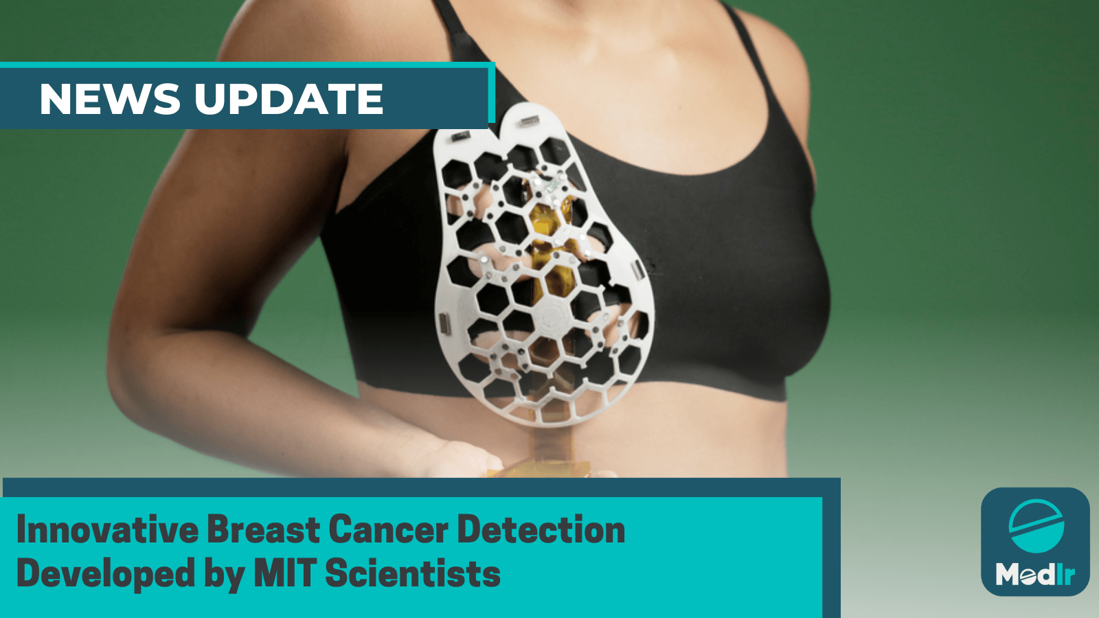
Massachusetts Institute of Technology scientists have developed a flexible patch for high-risk patients who may need more than recommended mammograms. The patch can take ultrasound images similar to medical centers' scans and fit into a bra. Interval cancers, which account for 20-30% of breast cancer cases, are more aggressive and require early detection. The development was published in Scientific Advances on July 28.
Canan Dagdeviren, an associate professor in MIT’s Media Lab and the senior author of the study, said in a release -
“We changed the form factor of the ultrasound technology so that it can be used in your home. It’s portable and easy to use, and provides real-time, user-friendly monitoring of breast tissue.”
Dagdeviren created a tiny ultrasound scanner using piezoelectric material, inspired by her late aunt's breast cancer. The flexible 3D-printed patch with honeycomb-like openings allows users to take images anytime. When combined with a matching bra, the scanner can be moved to six different spots, enabling breast imaging without special training.
To assess its effectiveness, the researchers tested the device on a 71-year-old individual with a history of breast cysts. The results were promising as the device detected cysts as small as 0.3 centimeters in diameter, up to a depth of 8 centimeters in the tissue, while maintaining a resolution comparable to traditional ultrasounds.
The team plans to create a mini, phone-sized imaging system for high-risk individuals to view images in the comfort of their homes. This could benefit patients without regular screening access and provide a convenient alternative to imaging centers-style ultrasound devices.
Study author Catherine Ricciardi, nurse director at MIT’s Center for Clinical and Translational Research, said in the release -
“Access to quality and affordable health care is essential for early detection and diagnosis. As a nurse I have witnessed the negative outcomes of a delayed diagnosis. This technology holds the promise of breaking down the many barriers for early breast cancer detection by providing a more reliable, comfortable, and less intimidating diagnostic.”